Skip to content

Treatment

Varicocele Embolization

Varicocele embolization is a minimally invasive, image-guided treatment for varicoceles — no scrotal incision, no general anesthesia, same-day discharge, and outcomes equivalent to surgery.

Varicocele embolization venogram with coils sealing the internal spermatic vein through a pinhole catheter, no incision
~90%
technical success rate of varicocele embolization
Per published SIR registries
60–70%
of infertile men see improved semen parameters within 6 months
Per published meta-analyses
~30–40%
spontaneous pregnancy rate within 12 months for couples with male-factor infertility
Per published cohorts
<5%
rate of post-procedure hydrocele (vs. ~7–15% with surgical ligation)
Per comparative studies

What it is

Varicocele embolization is an outpatient, image-guided procedure that treats a varicocele — the abnormally enlarged pampiniform plexus of veins draining the testicle — without a scrotal incision. Under live X-ray (fluoroscopic) guidance, a small catheter is advanced through a single pinhole in a neck or groin vein into the dilated internal spermatic (gonadal) vein. The refluxing vein is sealed with platinum coils, sometimes paired with a sclerosing agent, so blood naturally reroutes through healthy collateral veins. The testicle continues to drain normally — only the abnormal refluxing pathway is closed. Because there is no scrotal cutdown, no general anesthesia, and no division of the spermatic cord, the testicular artery, lymphatics, and vas deferens are never at risk. Published series report technical success of roughly 90% and pregnancy and semen-parameter outcomes that match or exceed microsurgical varicocelectomy, with markedly lower hydrocele rates and a fraction of the recovery time. Dr. Rastinehad has performed hundreds of varicocele embolizations and teaches the technique nationally.

Best suited for

  • Men with symptomatic varicoceles (scrotal pain, heaviness, or swelling worse with standing).
  • Couples with male-factor infertility and abnormal semen analysis (count, motility, or morphology) linked to a clinical or sub-clinical varicocele.
  • Adolescents and young men with documented testicular volume loss (>20% asymmetry) on the affected side.
  • Patients with recurrent or persistent varicocele after prior surgical ligation or embolization.
  • Bilateral varicoceles — both sides treated in a single session through one access site.
  • Anyone seeking a no-incision, no-general-anesthesia alternative to microsurgical varicocelectomy.

How it works

  1. 1Local anesthesia at a small puncture in the right internal jugular vein (neck) or right femoral vein (groin).
  2. 2A diagnostic venogram confirms the anatomy and identifies all refluxing channels, including collaterals that surgery cannot see.
  3. 3A microcatheter is navigated through the inferior vena cava into the left (or right) internal spermatic vein.
  4. 4Platinum coils, sometimes combined with a sclerosing agent, are deployed to permanently close the abnormal vein.
  5. 5The catheter is removed and the access site closed with a small adhesive dressing. No stitches.

Varicocele embolization, step by step

Neck access, catheter advanced into the spermatic vein, platinum coil embolization, varicocele decompresses.

  1. 1. Jugular vein access

    A small puncture is made in the right internal jugular vein in the neck under local anesthesia. No scrotal incision, no scalpel, and no general anesthesia.

  2. 2. Microcatheter navigation

    Under live X-ray guidance, a thin microcatheter is steered through the vena cava and into the refluxing left (or right) internal spermatic vein. A diagnostic venogram confirms every abnormal channel, including collateral pathways that surgery cannot directly visualize.

  3. 3. Coil deployment

    Platinum coils, sometimes combined with a sclerosing agent, are placed to permanently close the abnormal refluxing vein. Healthy collateral veins continue to drain the testicle normally.

  4. 4. Varicocele decompresses

    With the refluxing pathway sealed, the varicocele decompresses. Scrotal heaviness and pain typically improve within 1–2 weeks, scrotal temperature normalizes, and semen parameters improve over 3–6 months.

Benefits

  • No incision, no scalpel, no sutures — small adhesive dressing only.
  • No general anesthesia required; light IV sedation and local numbing.
  • Faster recovery than open or laparoscopic ligation (back to desk work in 1–2 days, vs. 1–2 weeks for surgery).
  • Equivalent or better success rates than surgical ligation in published series.
  • Bilateral varicoceles treated in the same single session through one access site.
  • Lower risk of hydrocele formation (<5%) compared with surgical ligation (7–15%).
  • Testicular artery, vas deferens, and lymphatics are never at risk — they are not in the vein being treated.
  • Both testicles continue to drain normally — only the abnormal vein is closed.

Possible risks

  • Mild bruising or tenderness at the small access site (neck or groin).
  • Brief lower-back or flank discomfort for 1–2 days (related to the embolic agent).
  • Rare allergic reaction to iodinated contrast dye.
  • Recurrence or persistence (5–10%), sometimes requiring a repeat embolization.
  • Very rare coil migration (treated technique-dependently).
  • No clinically meaningful radiation dose for the average patient, though pregnancy is a contraindication.

Embolization vs. surgical alternatives

Microsurgical varicocelectomyGold-standard surgery; requires a 2–3 cm inguinal or sub-inguinal incision, general or spinal anesthesia, 1–2 weeks off work, and carries a small risk of hydrocele (~7%), testicular artery injury, or wound complication. Slightly lower recurrence (~1–2%) than embolization but at the cost of a true operation.
Laparoscopic ligationRequires general anesthesia and three abdominal port incisions. Longer recovery, higher hydrocele rate (~10%), and a small risk of bowel or vascular injury. Rarely first-line today.
Open ligation (Palomo / retroperitoneal)Highest recurrence (~15%) and hydrocele rates of any approach. Largely supplanted by microsurgery and embolization.
Embolization (this procedure)No incision, no general anesthesia, same-day discharge, 1–2 day recovery. Bilateral disease treated in one session. Recurrence 5–10%, hydrocele <5%. Equivalent fertility and pregnancy outcomes to microsurgery in published comparative series.
ObservationReasonable for asymptomatic varicoceles with normal semen analysis and no testicular volume loss. Re-evaluate annually with exam and (if relevant) semen analysis.

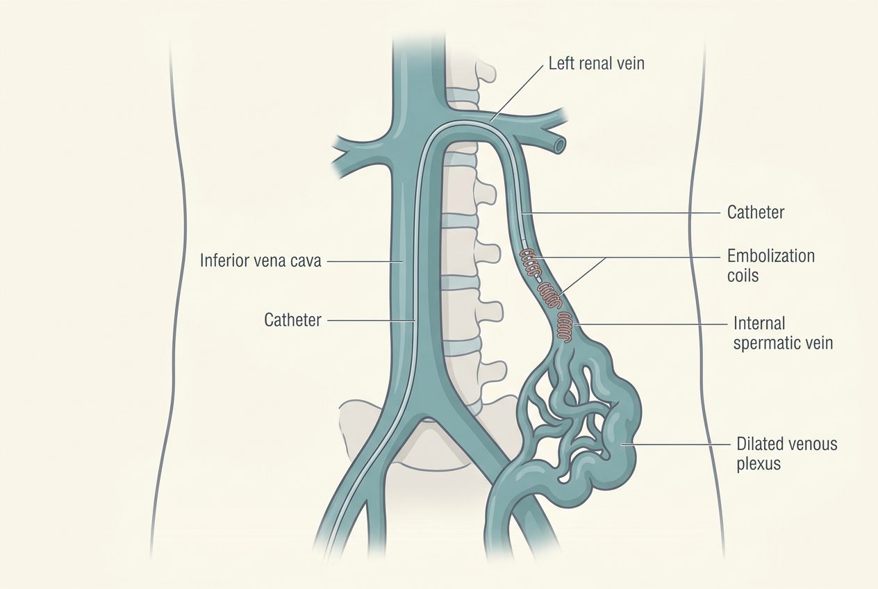
Why varicoceles affect fertility and testicular health

The testicles sit outside the body because sperm production (spermatogenesis) requires a temperature 2–4°C below core body temperature. The pampiniform plexus — a fine network of veins surrounding the testicular artery in the spermatic cord — acts as a heat exchanger, cooling arterial blood before it reaches the testicle. In a varicocele, valves in the internal spermatic vein fail, blood pools and refluxes downward, and the plexus dilates. The heat-exchange mechanism breaks down and intra-testicular temperature rises.

The result is a measurable cascade: heat stress, oxidative damage from reactive oxygen species, sperm DNA fragmentation, impaired Sertoli and Leydig cell function, and over time, testicular volume loss. This is why a varicocele is the single most common correctable cause of male-factor infertility — it is found in roughly 40% of men with primary infertility and 80% of men with secondary infertility. Treating the varicocele removes the reflux, restores normal scrotal temperature, and gives the testicle months to recover. Sperm take about 74 days to mature, so the first meaningful semen analysis after embolization is at 3 months, with continued improvement out to 6–12 months.

Why most varicoceles are on the left

Anatomy explains the laterality. The left internal spermatic vein drains at a 90° angle into the left renal vein, a longer and higher-pressure pathway than the right, which drains obliquely into the inferior vena cava. The left side is also subject to the 'nutcracker' compression of the renal vein between the aorta and superior mesenteric artery. About 85% of varicoceles are left-sided, 10% are bilateral, and isolated right-sided varicoceles are rare enough that they warrant an imaging look for a retroperitoneal cause.

Embolization is uniquely well-suited to this anatomy. The same catheter that maps the refluxing vein can also identify and treat collateral channels — periureteric, retroperitoneal, or cross-pelvic veins — that a scrotal-side surgery cannot directly see. These hidden collaterals are a major reason varicoceles recur after surgical ligation, and addressing them in the same session is one of the technical advantages of the image-guided approach.

Embolization vs. microsurgery: how to choose

Both microsurgical varicocelectomy and embolization are guideline-supported, evidence-based options. The right answer depends on anatomy, goals, and personal preference. Choose embolization when avoiding general anesthesia, returning to work quickly, treating bilateral disease in one session, or treating a recurrence after prior surgery is a priority. Choose microsurgery when a hydrocele or scrotal exploration is already on the table, when access to interventional radiology is limited, or when published recurrence rates of 1–2% (vs. 5–10% with embolization) are decisive for the patient.

  • Anesthesia: local + sedation (embolization) vs. general or spinal (surgery).
  • Recovery: 1–2 days (embolization) vs. 1–2 weeks (surgery).
  • Hydrocele risk: <5% (embolization) vs. 7–15% (surgery).
  • Bilateral disease: one session, one access site (embolization) vs. two incisions (surgery).
  • Recurrence: 5–10% (embolization) vs. 1–2% (microsurgery).
  • Fertility and pregnancy outcomes: equivalent in published comparative series.

What the day of your procedure looks like

Plan for about half a day. You arrive 60–90 minutes before the scheduled time for check-in, an IV, and a brief meeting with Dr. Rastinehad and the anesthesia team. The procedure itself runs 45–90 minutes — local anesthesia at the neck or groin access, gentle sedation, live X-ray imaging of the spermatic vein, and coil deployment. You will not have a urinary catheter and there is no scrotal dressing.

Recovery takes 1–2 hours in a comfortable observation bay. Most patients eat, walk to the bathroom, and are driven home the same afternoon. The first 24–48 hours typically include mild flank or lower-back ache (a normal response to the embolic material) that responds to ibuprofen or acetaminophen, plus a small bruise at the access site. By day 2 most men are back at a desk job; by week 2 they are back to the gym, lifting, and intercourse.

Cost, insurance coverage, and what to expect on billing

Varicocele embolization is FDA-cleared, has been covered by Medicare for decades, and is covered by virtually every major commercial insurer when performed for a clinically appropriate indication — symptomatic varicocele, abnormal semen analysis with planned conception, or testicular volume loss in an adolescent. Pre-authorization typically requires documentation of a clinical varicocele on exam, a confirming scrotal Doppler ultrasound, and, when applicable, a recent semen analysis.

The office handles benefits verification and pre-authorization before scheduling. Out-of-pocket cost depends on your plan's deductible, coinsurance, and out-of-network status — you will get a written estimate in advance, not a surprise. For self-pay patients, the all-in cost is generally substantially lower than microsurgical varicocelectomy because there is no hospital stay, no general anesthesia, and no operating-room fee.

Preparing for your procedure

  • Send recent scrotal ultrasound and (if applicable) semen analysis for review.
  • Hold blood thinners as directed (typically 2–5 days; many can continue uninterrupted).
  • Light meal in the morning; clear liquids only 2 hours before.
  • Arrange a ride home; no driving for 24 hours after sedation.
  • Bring a supportive undergarment (athletic supporter or snug briefs) for comfort the first 24 hours.

Why patients choose Dr. Rastinehad

Dr. Rastinehad is dual-trained in urology and interventional radiology and has performed hundreds of varicocele embolizations. His urologic background means he understands the fertility, hormonal, and pain considerations driving treatment, not just the catheter work — your evaluation, the procedure itself, and your fertility follow-up are all delivered by the same physician.

Used to treat

Conditions Dr. Rastinehad treats with Varicocele Embolization.

FAQ

About Varicocele Embolization

Answers patients most commonly ask before their consultation.

60–70% of men with male-factor infertility see improvements in sperm count, motility, or morphology within 3–6 months. Spontaneous pregnancy rates of 30–40% within a year are reported in published series, comparable to microsurgical varicocelectomy.

Medically reviewed by Art Rastinehad, D.O. — board-certified urologist and interventional radiologist, Lenox Hill / Northwell Health. This page is for general education and is not a substitute for medical advice.

Ready to talk?

Personalized urologic care begins with a conversation.

Schedule a consultation to review your imaging, lab work, and treatment options.